28+ Wahrheiten in Loculated Pleural Effusion Meaning: Care guide for pleural effusion.. Send aspirated fluid for cytology. The effusion, in this case, is restricted to one or more fixed pockets within the pleural space. Pleural effusion that is confined to one or more fixed pockets in the pleural space. While breathing, when the chest moves, the lining also moves along with it smoothly within the chest cavity to let the lung expand and inhale air. Occasionally, a focal intrafissural fluid collection may look like a lung mass.
Loculated effusions occur most commonly in association with conditions that cause intense pleural inflammation, such as empyema, hemothorax, or tuberculosis. They may result from a variety of pathological processes which overwhelm the pleura's ability to reabsorb fluid. Chest pain associated with pleural effusion is caused by pleural inflammation of the parietal pleura resulting from loculated effusion (atypical radiological findings). This is most likely related to infection unless a trauma has recently occurred and then this can be related to secondary infection of. A pleural effusion occurs either because of an imbalance between the osmotic and cough, if present, in a patient with a pleural effusion, usually means that there is something affecting the small effusions, whether loculated or not, will not be expected to cause tracheal deviation.
Consolidation and collapse in the right lung with a large ... from www.researchgate.net Pleural effusion (transudate or exudate) is an accumulation of fluid in the chest or on the lung. Meaning of loculated pleural effusion medical term. If none is present the fluid is virtually always a transudate. Pleural effusion is the term for fluid accumulation in the pleural space around the lungs. Treatment depends on the cause. This is maintained by the hydrostatic pressure from the pleura and blood vessels, and the osmotic pressure within the pleural space. Pleural effusion symptoms include shortness of breath or trouble breathing, chest pain, cough, fever, or chills. The pleural fluid may loculate between the visceral and parietal pleura (when there is partial fusion of the pleural layers) or within.
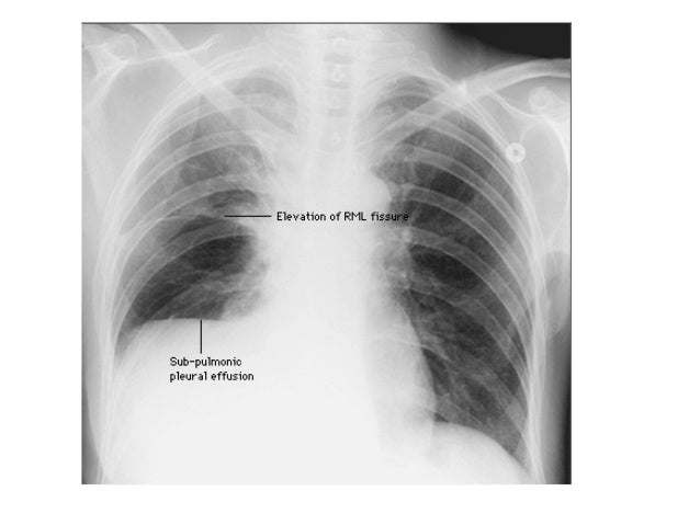
This situation most commonly is seen in patients with heart failure.
While breathing, when the chest moves, the lining also moves along with it smoothly within the chest cavity to let the lung expand and inhale air. Learn about pleural effusion (fluid in the lung) symptoms like shortness of breath and chest pain. Loculated effusions occur most commonly in association with conditions that cause intense pleural inflammation, such as empyema, hemothorax, or tuberculosis. Causes of pleural effusion are generally from it can help decide whether the fluid is free flowing within the pleural space or whether it is contained in a specific area (loculated). Occasionally, a focal intrafissural fluid collection may look like a lung mass. Multiloculated means that the fluid isn't just one single continuous collection but loculated pleural: Pleural effusions are abnormal accumulations of fluid within the pleural space. There is normally a tiny amount of fluid between the two layers of pleura. They may result from a variety of pathological processes which overwhelm the pleura's ability to reabsorb fluid. Pleural effusions accompany a wide variety of disorders of the lung, pleura, and systemic disorders. Pleural effusion that is confined to one or more fixed pockets in the pleural space. Loculated pleural effusion masquerading as mediastinal tumour had been reported but pleural effusion that conformed to the contour of a lung lobe is rare. More than one half of these massive pleural effusions are caused by malignancy;
Pleural effusion is classically divided into transudate and exudate based on the light criteria. A pleural effusion is accumulation of excessive fluid in the pleural space, the potential space that surrounds each lung. Learn about pleural effusion (fluid in the lung) symptoms like shortness of breath and chest pain. Pleural fluid/serum ldh ratio >0.6. There is normally a tiny amount of fluid between the two layers of pleura.
2 Lung Ultrasound Pre-Reading for FCUS course - Intensive ... from intensivecarenetwork.com More than one half of these massive pleural effusions are caused by malignancy; Pleural effusion is fluid buildup in the space between the layers of the pleura. Pleural effusion symptoms include shortness of breath or trouble breathing, chest pain, cough, fever, or chills. Multiloculated means that the fluid isn't just one single continuous collection but loculated pleural: Loculated effusions are collections of fluid trapped by pleural adhesions or within pulmonary fissures. Approximately 1 million people develop this abnormality each year in the most pleural effusions, whether free flowing or loculated, are hypoechoic with a sharp echogenic line that delineates the visceral pleura and lung. Recent reports have advocated the use of. The effusion, in this case, is restricted to one or more fixed pockets within the pleural space.
The pleura is a thin membrane that lines the surface of your lungs and the inside of your chest wall.
A pleural effusion is accumulation of excessive fluid in the pleural space, the potential space that surrounds each lung. While breathing, when the chest moves, the lining also moves along with it smoothly within the chest cavity to let the lung expand and inhale air. Pleural effusions accompany a wide variety of disorders of the lung, pleura, and systemic disorders. Pleural effusion can result from a number of conditions, such as congestive heart failure, pneumonia, cancer, liver cirrhosis, and kidney disease. Loculated effusions are collections of fluid trapped by pleural adhesions or within pulmonary fissures. Treatment depends on the cause. They may result from a variety of pathological processes which overwhelm the pleura's ability to reabsorb fluid. There is normally a tiny amount of fluid between the two layers of pleura. When a person has pleural effusion, it means that fluid has collectedtrusted source in the space between their lungs and chest cavity, or pleural the lungs and the chest cavity both have a lining that consists of pleura, which is a thin membrane. Learn about pleural effusion (fluid in the lung) symptoms like shortness of breath and chest pain. Understanding pleural effusion pleura refers to thin membranes that line the lungs and the inside of the chest cavity. Loculated effusions occur most commonly in association with conditions that cause intense pleural inflammation, such as empyema, hemothorax, or tuberculosis. A pleural effusion is a collection of fluid next to the lung.
Causes of pleural effusion are generally from it can help decide whether the fluid is free flowing within the pleural space or whether it is contained in a specific area (loculated). When a person has pleural effusion, it means that fluid has collectedtrusted source in the space between their lungs and chest cavity, or pleural the lungs and the chest cavity both have a lining that consists of pleura, which is a thin membrane. Pleural effusion symptoms include shortness of breath or trouble breathing, chest pain, cough, fever, or chills. The pleura are thin membranes that line the lungs and the inside of the chest cavity and act to lubricate and facilitate breathing. Pleural effusion that is confined to one or more fixed pockets in the pleural space.
Diagnostic Imaging of Pleural Lesions from image.slidesharecdn.com Pleural fluid/serum protein ratio >0.5. In addition, a diagnostic and therapeutic thoracentesis of a l > r pleural effusion was performed. Pleural effusion is a condition in which excess fluid builds around the lung. Learn vocabulary, terms and more with flashcards, games and other study tools. Pleural fluid ldh > two thirds of upper limit for serum ldh. When you have a pleural effusion, fluid builds up in the space between the layers of your pleura. In healthy lungs, these membranes ensure that a. This is most likely related to infection unless a trauma has recently occurred and then this can be related to secondary infection of.